


MCMASTER: McMaster researchers found that bacteriophages treated under specific conditions form flower-like structures that are highly efficient in targeting bacteria, opening new possibilities for the treatment and detection of diseases.
A team of McMaster researchers who routinely work with bacteriophages – viruses that eat bacteria – made a remarkable discovery while preparing slides to view under a powerful microscope.
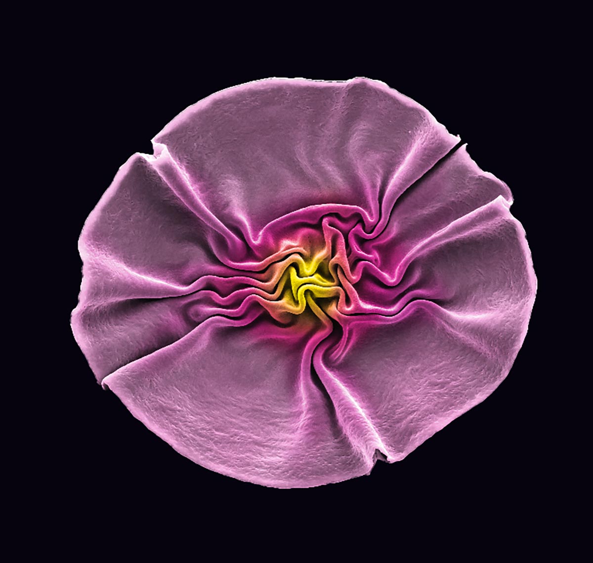
After treating samples of bacteriophages, informally known as phages, to view them alive under an electron microscope, the researchers were surprised to find that they had formed into three-dimensional shapes resembling sunflowers, but only two-tenths of a millimeter across.
With a little prompting, nature had produced the very type of structure that experts in the field had been attempting to artificially create for decades—structures that proved to be 100 times more efficient than unlinked phages at finding elusive bacterial targets.
According to the researchers, the ability to create such structures opens up possibilities for the detection and treatment of many forms of disease, all using natural materials and processes.
Their findings are detailed in a newly published article in the journal Advanced Functional Materials.
Unveiling Unique Viral Forms
The initial discovery was a happy accident flowing from everyday laboratory work.
Rather than expose the sample phages to typical preparation processes, which involve temperatures or solvents that kill viruses, lead author Lei Tian and his colleagues elected to treat them with high-pressure carbon dioxide instead. Tian, now a principal investigator at Southeast University in China, led the research while he was a PhD student and later a post-doctoral research fellow at McMaster.
While the researchers are used to seeing the microscopic viruses do amazing things, after the treatment they were stunned to see the phages had grouped together in such complex, natural, and very useful forms.
“We were trying to protect the structure of this beneficial virus,” Tian says. “That was the technical challenge we were trying to overcome. What we got was this amazing structure, which was made by nature itself.”
The researchers captured images of the formations using the facilities of the Canadian Centre for Electron Microscopy, located at McMaster, and spent the last two years unlocking the process and showing how the new structures can serve very useful purposes in science and medicine.
“It was an accidental discovery,” says the paper’s corresponding author Tohid Didar, a mechanical engineer who holds the Canada Research Chair in Nano-biomaterials. “When we took them out of the high-pressure chamber and saw these beautiful flowers, it completely blew our minds. It took us two years to discover how and why this happened and opened the door to being able to create similar structures with other protein-based materials.”